Showing posts with label General Medicine. Show all posts

Tuesday, January 10, 2012

Fever with rash

A male patient of 20 years presented to emergency with complaints of fever and altered sensorium of 1 day duration. On examination this rash was present. What is the diagnosis?


a) Purpura fulminans
b) Rocky Mountain Spotted Fever
c) Sterptococcal cellulitis
d) Dengue Fever


Answer is a) Purpura Fulminans ( Severe Meningococcal septic rash)

Massive intracutaneous hemorrhages are apparent in a patient with meningococcemia. The bleeding is related in part to thrombocytopenia, which is often part of the syndrome of disseminated intravascular coagulation, and in part to vascular damage caused by the meningococci, which can often be found invading the blood vessels. The lesions shown here are sometimes referred to as “purpura fulminans” and are associated with a high mortality rate.

Read more about Meningococcemia HERE

Monday, January 9, 2012

Colonoscopy

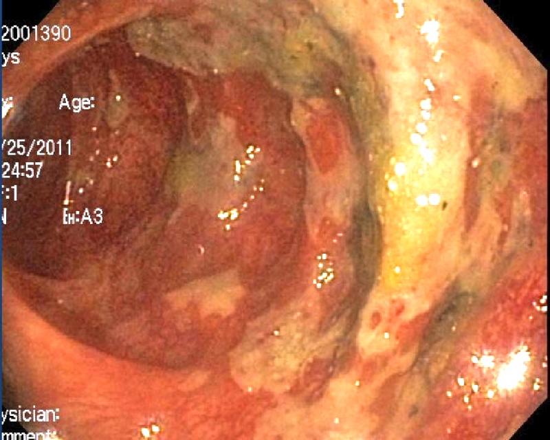
An 61 year old patient had urinary tract infection. He received antibiotic therapy. He presented later with diarrhea. Colonoscopy was done photograph is given below. What is the Diagnosis?


a) Campylobacter
b) Amebiasis
c) Pseudomembranous colitis
d) Cryptosporidiosis
e) Viral GE


Answer is c) Pseudomembranous colitis

This is classical appearence on colonoscopy. The classic appearance of 2-mm to 10-mm raised yellow nodules is pathognomonic. These lesions are usually discrete but may become confluent plaques in more advanced cases. Usually colonoscopy is not done as it is invasive procedure. Diagnosis is usually made by detecting C.difficale toxins.


Read More aout the condition HERE

Sunday, January 8, 2012

Diagnose It

A 40 year female having swelling of right foot since 11 years. The nodules on the swelling were painful, drained periodically and made weight bearing difficult. She had received courses of antibiotics with partial responses. What is the diagnosis?


a) Tuberculous osteomyelitis
b) Blastomycosis
c) Madura foot
d) Histoplasmosis
e) Leishmaniasis


Answer is c) Madura foot.

Examination showed soft-tissue swelling, multiple red, boggy, subcutaneous nodules, and draining sinus tracts. Culture of a specimen from a punch biopsy failed to grow any organisms. However, histologic analysis revealed whitish-yellow granules and the presence of non–acid-fast filamentous bacteria consistent with Madura foot. This condition can be caused by either filamentous actinobacteria (actinomycotic mycetoma) or true fungi (eumycotic mycetoma). The pale color of the granules combined with the fact that the patient was from Mexico, where most mycetomas are caused by actinomadura species, made actinomycotic mycetoma the more likely diagnosis. Magnetic resonance imaging of the foot was performed, and the scan obtained indicated possible osteomyelitis.

Read more about madura foot HERE

Saturday, January 7, 2012

MRI Brain

What is the diagnosis?


a) Cerebra Aneurysm
b) Chiari I malformation
c) Neurofibramatosis
d) Pagets Disease
e) Subdural Hematoma


Answer is b) Chiari I Malformation

The T1-weighted sagittal image shows a tonsillar herniation (type I Chiari malformation) more than 5 mm below the level of the foramen magnum

Read more about Arnold Chiari Malformations HERE